Mission Statement: The innate immune system in retinal degenerative diseases – from understanding to novel innovative therapies
Loss of vision due to inherited (e.g. retinitis pigmentosa) or multifactorial (e.g. age-related macular degeneration) retinal disease is a devastating and in most cases unstoppable prognosis and accompanied by large constraints in the life quality of affected patients. To date, the Retnet database (https://sph.uth.edu/retnet/) lists a total of almost 300 genes and loci linked with retinal degenerations. 25% of these genes are causative for the most frequent monogenic group of disorders, retinitis pigmentosa (RP). On the other hand, the leading cause of legal blindness in the elderly of industrialized nations today is age-related macular degeneration (AMD), a complex genetic disease. In contrast to RP, a multitude of genetic risk variants as well as environmental risk factors contribute to the development of AMD and ultimately loss of vision.
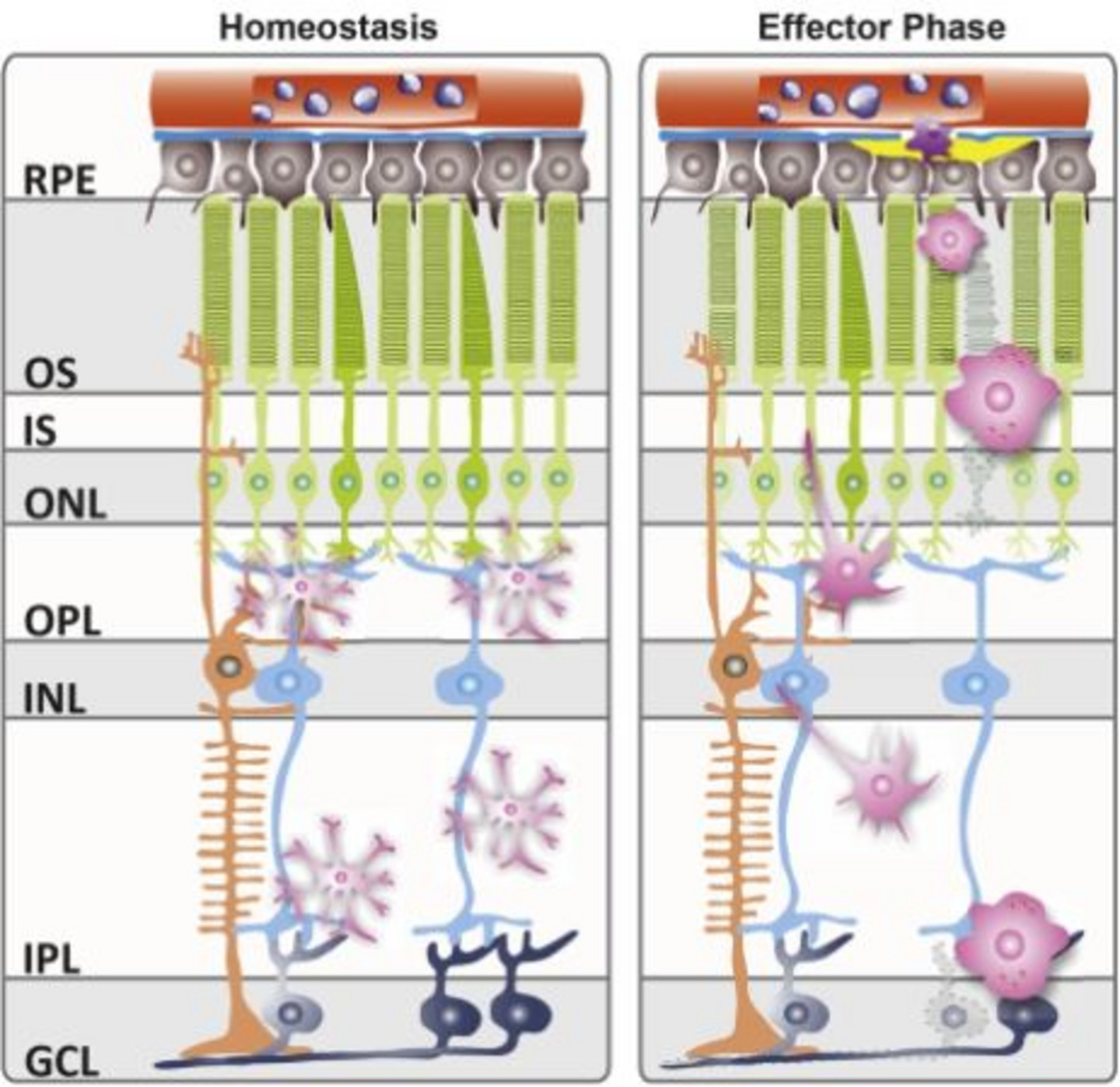
Microglial cells are the resident phagocytes of the central nervous system (CNS), including the retina, and play pivotal roles in innate immune responses and regulation of homeostasis in the healthy and degenerating CNS. Reactive microgliosis is a common hallmark of neurodegenerative diseases and chronic pro-inflammatory microglial reactivity negatively contributes to disease progression (Langmann, 2007). Previous research from our laboratory could demonstrate the important role of innate immune activation during early retinal pathogenesis (Weigelt et al., 2007). Interestingly, we and others could show that this early reactivity of microglial cells is broadly independent of the underlying genetic defect or cause of retinal degeneration (Karlstetter et al., 2010a, Ebert et al., 2009, Karlstetter et al., 2014b, Scholz et al., 2015b)
Our laboratory therefore postulates that targeted microglia-directed immunotherapy could represent an early and feasible strategy to attenuate progression of a variety of retinal degenerative diseases (Karlstetter et al., 2015). Using various cell culture and animal models together with modern tools of molecular biology, we constantly aim to identify novel microglial activation biomarkers and suitable target structures for microglia-directed immunotherapy in degenerative diseases of the retina.
Our previous in vivo and in vitro analyses enabled us to identify and characterize two novel biomarkers for microglial reactivity in the retina: Activated Microglia Whey Acidic Protein (AMWAP) and the Translocator Protein (18 kDa) (TSPO) (Karlstetter et al., 2010b, Aslanidis et al., 2015, Karlstetter et al., 2014a). Further, we could demonstrate successful therapeutic microglia modulation and neuroprotection in mouse models of hereditary and light-induced retinal degeneration using the natural compounds curcumin and docosahexaenoic acid (DHA), the antibiotic minocycline as well as Interferon-ß and a synthetic TSPO ligand (Ebert et al., 2009, Mirza et al., 2013, Scholz et al., 2015b, Luckoff et al., 2016, Scholz et al., 2015a).
By identifying, characterizing and therapeutically utilizing further key molecules of microglial reactivity during retinal degeneration we aim to contribute to the development of promising novel therapeutic approaches for the treatment of retinal inflammation and degeneration.
Translocator protein 18 kDa (TSPO)/ anti-VEGF combination therapy in Age-Related macular degeneration (AMD)
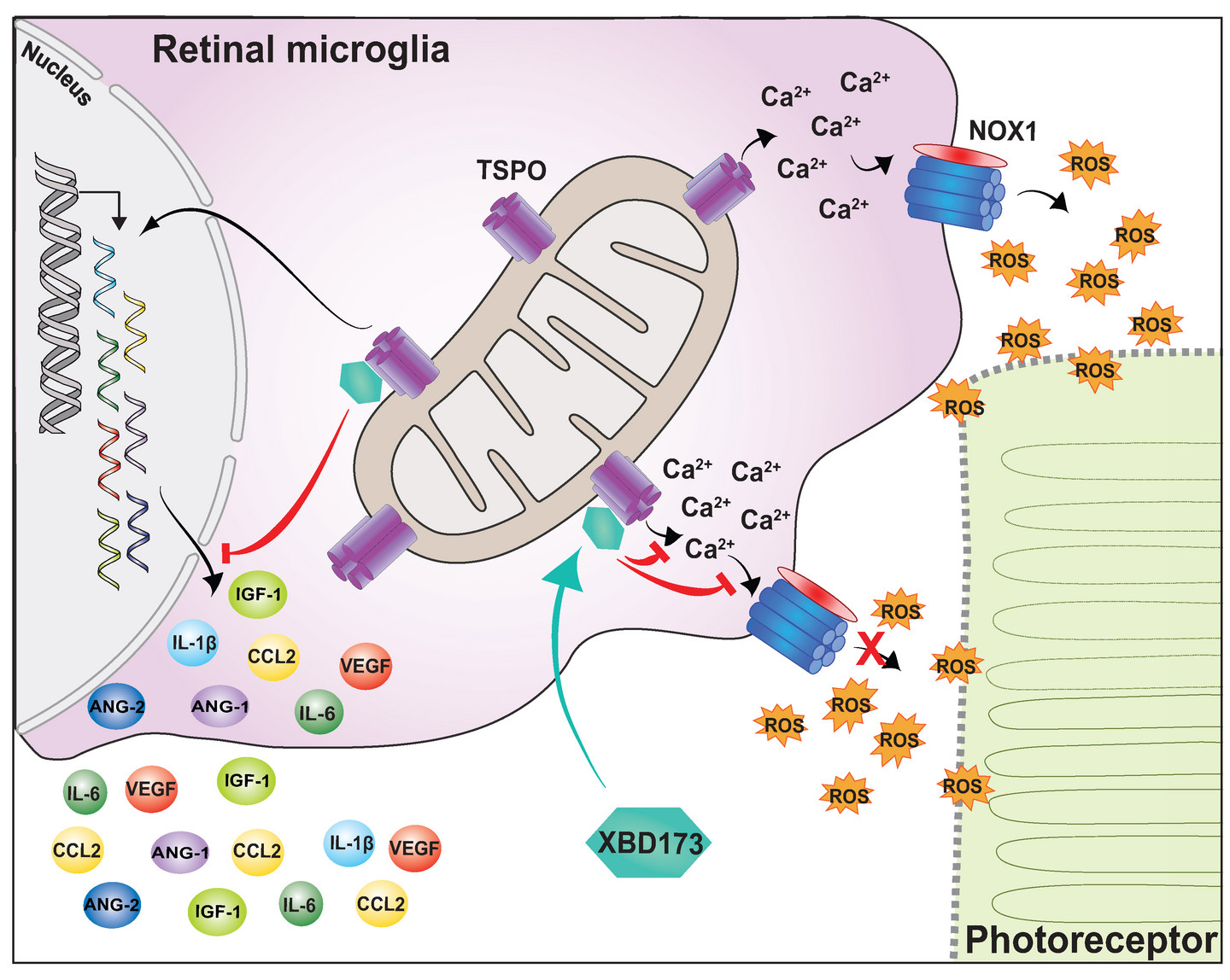
The innate immune system is one of the relevant mechanisms in the development and progression of age-related macular degeneration (AMD). With the translocator protein (18 kDa) (TSPO), our research group discovered a new regulator of microglia activation in the eye. TSPO controls the release of neurotoxic oxygen radicals (ROS) in the retina by modulating the Ca2 + content and the NADPH oxidase 1 enzyme (NOX1) of the immune cells. Genetic deficiency of TSPO in microglia or therapy attempts via the TSPO ligand XBD173 showed a strong reduction in microglia and a reduced formation of new vessels in the laser CNV mouse model of wet AMD. In the planned project, the expression of TSPO in immune cells from donor retinas of AMD patients and controls will be compared. Next, a combination therapy of anti-VEGF drugs with the TSPO ligand XBD173 will be investigated in the laser-CNV mouse model. Since this novel approach addresses both the angiogenesis and the damaging immune response in the eye, we expect a significant synergistic outcome. These project data could potententially constitute a first step in investigating an immunomodulatory/anti-angiogenic combination therapy for wet AMD.
Translocator protein 18 kDa (TSPO) in microglia reactivity and immunotherapy
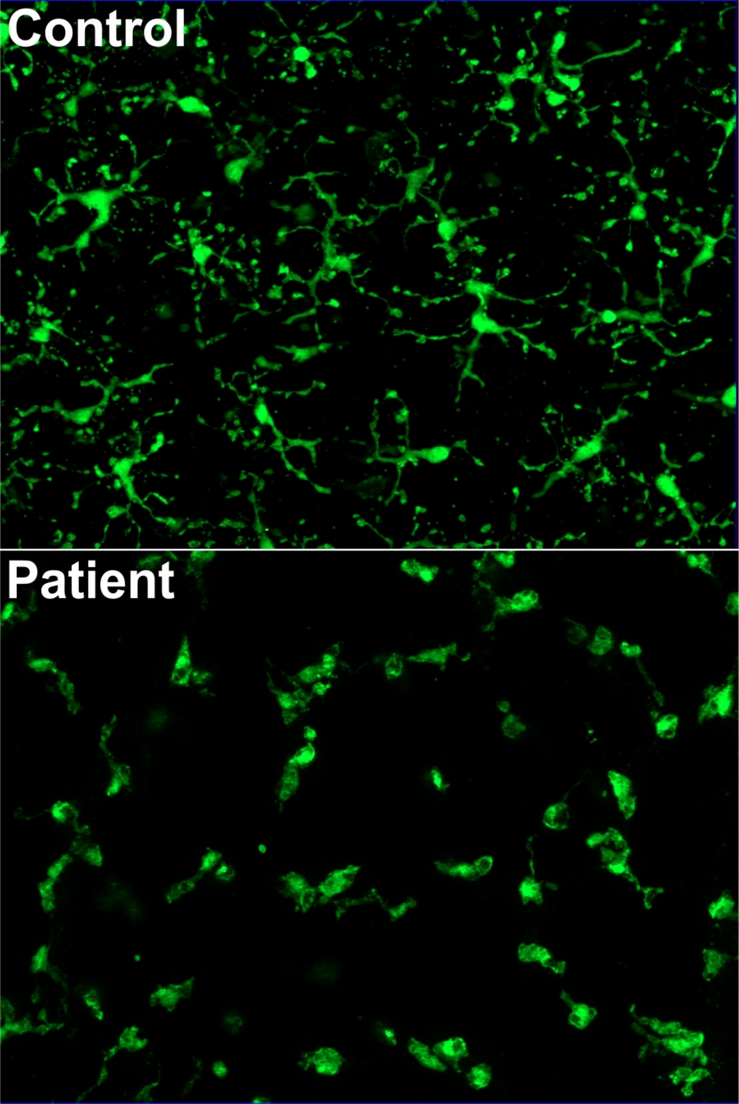
The translocator protein (18 kDa) (TSPO) is a mitochondrial protein expressed in reactive glial cells and a biomarker for gliosis in the brain but has just recently entered the spotlight in retinal degeneration research (Karlstetter et al., 2014a, Scholz et al., 2015a). We could show strong upregulation of TSPO transcript and protein levels in reactive microglial cells in vitro, microglia of the aSMase-deficient, retinoschisin-deficient and white light-induced retinal degeneration mouse models as well as TSPO expression in microglia of the human retina (Karlstetter et al., 2014a, Dannhausen et al., 2015, Scholz et al., 2015a). These were the first reports of TSPO as a microglial reactivity marker and potential therapeutic target in the retina.
Since TSPO is selectively upregulated in pathologies of the CNS, in recent years growing interest in the use of specific TSPO ligands as a strategy to attenuate neuroinflammation has emerged. In this regard, we could show that the synthetic TSPO ligand XBD173 effectively suppresses pro-inflammatory microglial reactivity in vitro by modulating pro-inflammatory microglial gene expression, migration, proliferation, NO secretion, neurotoxicity, proliferation, morphology and phagocytic capacity as well as reducing the number of amoeboid alerted microglia in organotypic murine retinal explant cultures (Karlstetter et al., 2014a).
In a following study we could, for the first time, demonstrate TSPO-mediated immunomodulatory and neuroprotective effects of XBD173 in mouse models of bright light-induced acute retinal degeneration. Specifically, systemic XBD173 treatment inhibited accumulation or reactive amoeboid microglia in the subretinal space and reduced both pro-inflammatory gene expression and photoreceptor apoptosis (Scholz et al., 2015a).
In summary, we contributed the first reports of TSPO as a microglial biomarker and potential therapeutic target to attenuate neuroinflammation in retinal degeneration.
Current TSPO-related projects in our laboratory focus on unraveling the microglia-specific regulation and induction of TSPO expression in retinal health and disease as well as on investigating the underlying molecular mechanisms of TSPO-mediated immunomodulation and neuroprotection using in vitro reporter assays as well as conditional microglia-specific knock-out mice and the laser-induced retinal injury model.
Investigating the role of microglia in the pathophysiology of diabetic retinopathy
Diabetic Retinopathy (DR) is a complication of diabetes and is a major cause of irreversible blindness among the working age population. DR has traditionally been regarded as a microvasculature disease, however mounting evidence from human patients and mouse models show that inflammation contributes significantly to the pathogenesis of DR. Inflammatory responses during retinal pathophysiology are orchestrated primarily by microglia cells, which constitute the resident immune cell population. However, the exact contribution of microglia to DR pathophysiology has remained unclear, largely due to lack of adequate DR animal models that fully mimic the human disease. Recently, it was demonstrated in two separate innovative mouse models that inhibition of platelet-derived growth factor (PDGF)-B/PDGF receptor beta (PDGFRβ) signaling during development of retinal vessels is sufficient to reproduce characteristic features of DR in adult mice. These novel mice models provide an essential tool for studying DR and development of novel therapies. Therefore in this project, we propose to perform an in-depth analysis of retinal microglia at different stages of DR. We also aim to fully characterize microglial roles in DR using i) microglial ablation with the oral PLX5622 ii) modulation of microglia reactivity with a second generation tetracycline, minocycline which we and others have shown to be a potent modulator of retinal microgliosis. This study will not only improve our understanding of microglia in DR pathogenesis but may also demonstrate microglia as potential therapeutic targets.
Colony stimulating factor 1 (Csf1) receptor blockade as novel tool to limit microglia reactivity in the light-damage model of retinal degeneration
Age-related macular degeneration (AMD) is a leading cause of blindness in industrialized countries. The etiology of AMD is not completely understood. However, genetic association studies in patient cohorts and in situ analyses in AMD eyes as well as animal models mimicking some features of AMD have identified an important contribution of dysregulated innate immunity in the eye. Using experimental and genetic models in mice we have previously established a potential pathogenic role for microglia and macrophages in the retina. In this project, we aim to evaluate the effects of a novel colony stimulating factor 1 (Csf1) receptor antagonist (PLX5622) on immune cell reactivity and retinal cell death in the light-damage model of retinal degeneration. In different microglia deactivation/depletion treatment regimens using oral PLX5622 before and after disease onset, we will focus on in vivo imaging of phagocytes in MacGreen reporter animals, their neurotoxic ROS secretion, and local complement factor expression using RNAscope in situ hybridization. These immune-related markers will be correlated with retinal cell death and degeneration in a temporal analysis of early and late phases after retinal injury. The results of these studies will provide new insights into the immune-related etiology of AMD and other retinal degenerative diseases and may foster the development of novel immunomodulatory treatments options.
Galectin-3 as a target for retinal immunomodulation
The innate immune system is one of the relevant pathomechanisms in the development and progression of age-related macular degeneration (AMD). Chronically activated microglial cells and its disturbed regulation contribute to retinal degeneration. The immune system therefore offers new therapy options for AMD. Our research group has identified polysialic acid from sugar biology as an endogenous protective function in the damaged retina. In the laser mouse model of wet AMD, we were able to demonstrate significant protection of the retina through immunotherapy with PolySia. In the planned project, a counterplayer of the immune-inhibiting polysia system, the pro-inflammatory galectin-3 sugar binding protein, is being investigated. Because, in contrast to wet AMD, there is no therapy option for dry AMD, this disease subtype will be the focus of our research. On the one hand, the harmful expression of galectin-3 in immune cells from donor retinas of AMD patients will be correlated with the course of the disease. On the other hand, we will test whether galectin-3 inhibition in the light damage model of dry AMD protects from retinal degeneration. Non-invasive optical coherence tomography (SD-OCT) and histological analyses of the retina are used for therapy monitoring. The reactivity of microglial cells will be determined both by histological stainings of sections and flat specimens and by molecular PCR markers. These new project data could be an important basis for initiating a possible clinical trial using Galectin-3 inhibitors for the treatment of dry AMD patients.
Microglial Interferon-ß signaling – road to AMD therapy?
Anti-VEGF is presently the gold standard in the treatment of choroidal neovascularization in wet AMD. However, inhibition of angiogenesis alone does not affect the cellular immunological events underlying CNV and also fails to be effective in the more common dry form of AMD. Furthermore, VEGF inhibition may cause a number of severe retinal and systemic adverse events. Therefore, there is an urgent need for identifying alternative approaches to treat AMD that is based on the underlying immunological pathogenesis. Type 1 Interferon signaling through the Ifnar receptor is a critical pathway in innate immune activity. Further, therapeutic interferon-β (IFN-β) has potent immunomodulatory effects on microglia both in vitro, in animal models of multiple sclerosis as well as being used for multiple sclerosis treatment in humans.
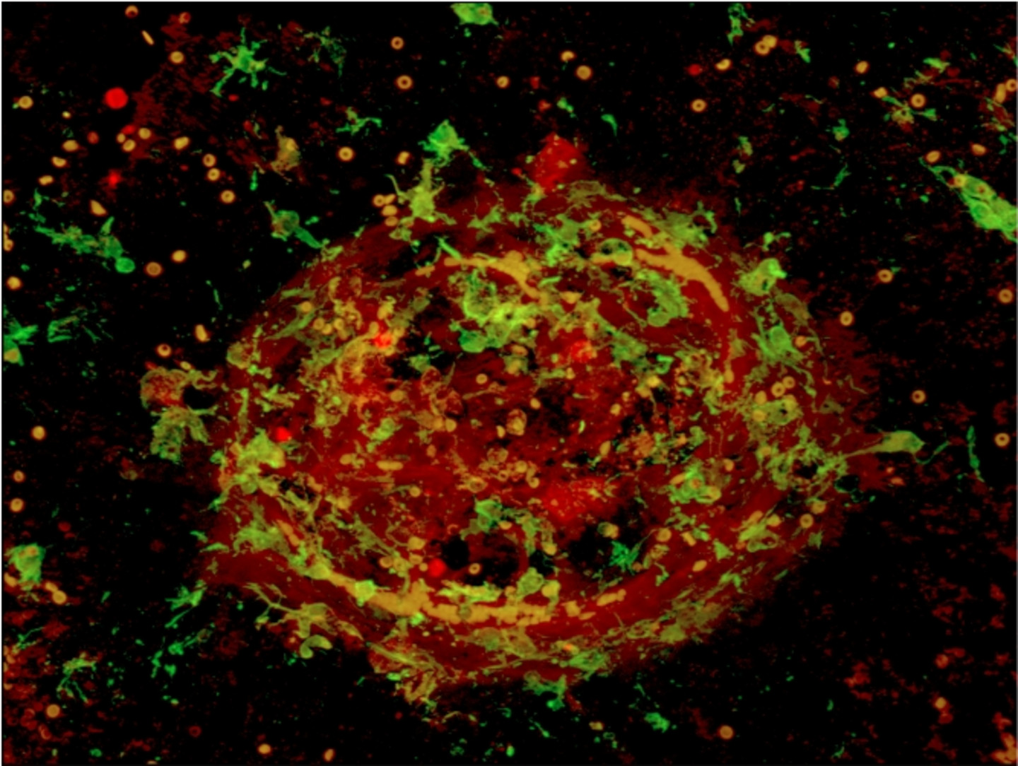
Our laboratory is interested in the role of IFN- β and its receptor Ifnar in the context of microglia in retinal degenerations like AMD. Using the laser-induced choroidal neovascularization (CNV) model of wet AMD, we could already demonstrate pivotal effects of Ifnar/IFN-β signaling in retinal microglia and macrophages. We could show an essential role of interferon-β signaling in regulating immune cell reactivity and pathological angiogenesis. Loss of Ifnar1 triggered microglia/macrophage activation, vessel leakage, and choroidal neovascularization (CNV). In contrast, IFN-β therapy attenuated retinal immune cell response and CNV development (Luckoff et al., 2016).Our findings indicate a key role for Ifnar signaling in retinal immune activation and the immunomodulatory potential of IFN-β as a promising new strategy for future therapy approaches to control chronic inflammation in AMD.
Polysialic acids as novel immunotherapeutics in AMD
Age-related macular degeneration (AMD) is the main cause of visual impairment and legal blindness in the industrialized world as more than one third of the population over the age of 75 develops AMD. Late-stage disease can either manifest as geographic atrophy or neovascular AMD. The neovascular or wet form is treated with intravitreally injected drugs targeting vascular endothelial growth factor (VEGF). In contrast, thereis currently no approved drug treatment for geographic atrophy, which is the atrophic or dry form of the disease.
AMD is associated with chronic innate immune activation specifically involving the complement system as well as activation of phagocytes and production of reactive oxygen species (ROS). Sialic acid polymers on the glycocalyx of healthy neurons inhibit complement activation and prevent ROS production of human phagocytes by acting on the inhibitory sialic-acid-binding immunoglobulin-like lectin-11 (SIGLEC11) receptor, but the therapeutic potential in AMD has not yet been evaluated. Using the humanized SIGLEC11 transgenic mouse model of laser-induced retinal damage, we investigate the therapeutic immunomodulatory potential of polysialic acids in age-related macular degeneration. Our experiments demonstrate potent prevention of retinal microglia and choroidal macrophage reactivity induced by laser coagulation. Furthermore, polysialic acids reduce retinal vascular leakage and deposition of the membrane attack complex (MAC) in this transgenic laser-damage animal model. These protective effects are mediated by two synergistic effects on the innate immune system: Firstly, polysialic acids prevent overt production and release of pro-inflammatory molecules from reactive phagocytes via human SIGLEC1. Secondly, polySia avDP20 directly interferes with activation of the alternative complement pathway.
These findings reveal modulation of inflammatory SIGLEC11 signaling by polysialic acid treatment as a novel promising therapy option for AMD, by inhibiting the damaging effects of innate immune activation.
The role of the complement system and microglia in AMD
Age-related macular degeneration (AMD) is a leading cause of blindness in industrialized countries. Early AMD manifests as drusen deposits between the outer retina and the pigment epithelium. AMD can progress to late stage forms including choroidal neovascularization (CNV) and an atrophic form with macular degeneration of photoreceptors and the pigment epithelium.
The etiology of AMD is not completely understood. However, genetic association studies in large patient cohorts and in situ analyses in AMD eyes as well as animal models mimicking some features of AMD have identified an important contribution of dysregulated innate immunity in the eye. Our project of the first funding period of FOR2240 has revealed local dysregulation of complement factors in ocular fluids of AMD patients and found a correlation of disease progression with hyperreflective foci (HF) as potential imaging markers for retinal immune cells. Furthermore, using a light damage paradigm and laser-triggered CNV in mice we established a comprehensive analysis of microglia and macrophages in the retina. Resident microglia-specific targeting of key immune pathways and immunomodulatory compounds could elucidate a vicious cycle of microglia reactivity, retinal degeneration and neoangiogenesis. These findings together provide evidence for an interaction of reactive microglia with aberrant complement proteins in the pathogenesis of AMD. However, the underlying mechanisms that link both systems are only incompletely explored. In this follow-up project we postulate that particularly the anaphylatoxin complement cleavage products C3a and C5a sustain chronic microglia reactivity via receptormediated recruitment and activation. We further postulate that hyperreflective foci could potentially mirror complement-regulated microglia responses and may therefore be used to monitor disease development in AMD patients and animal models. The project is subdivided into four specific aims in which we will determined the impact of C3a/C5a anaphylatoxins on microglia in vitro, test the effects of microglial C3a/C5a receptor deficiency in the laser-CNV and light damage models, evaluate the expression and localization of C3aR/C5aR in human retinal sections, and finally correlate the dynamics of hyperreflective foci with ocular complement factors in AMD patients and controls. The results of these studies will provide new insights into the immune-related etiology of AMD and may foster the development of novel effective treatments.

Function and pathophysiology of the retinitis pigmentosa gene FAM161A
Retinitis Pigmentosa (RP) describes a group of related hereditary retinal degenerative disorders, in which mutations in retina-specific genes cause loss of functional photoreceptors ultimately leading to blindness. Following a combined approach of homozygosity mapping and CRX ChIP-seq, we could previously identify loss-of-function mutations in the FAM161A gene as a cause for autosomal-recessive RP (Langmann et al., 2010). FAM161A expression is tightly regulated by the master photoreceptor transcription factor CRX as shown by organotypic reporter assays in explanted mouse retinas.
Functional studies by our group and collaborators revealed localization of FAM161A protein to the ciliary region, linking photoreceptor outer and inner segments, as well as important functions in microtubule-mediated intracellular protein trafficking (Zach et al., 2012). Accordingly, genetically modified mice lacking functional FAM161A protein show very early disorganization of photoreceptor outer segments accompanied by inflammatory microglial reactivity followed by complete loss of the light-sensitive photoreceptor cells by 6 months of age. Using high-resolution and electron microscopy, we could identify defects in the transportation of important photoreceptor proteins along the connecting cilium of photoreceptor cells as an underlying disease mechanism (Karlstetter et al., 2014b).
Our laboratory employs the valuable FAM161A-deficient ciliopathy model to gain further insight into photoreceptor function in health and disease as well as a tool for investigating novel gene-replacement and immunomodulatory therapies.